

In the microscopic world of molecular biology, every experiment is a journey into the unknown. Along this journey, probe‑based melting curve analysis offers a unique perspective, revealing the secrets of DNA sequences.
What is a Melting Curve?
First, let us clarify the basic concept of a melting curve. In real‑time quantitative PCR (qPCR) experiments, when double‑stranded DNA (dsDNA) is heated, the hydrogen bonds between complementary bases gradually break, separating the duplex into two single strands. This process is known as DNA “melting.” As temperature increases, the fluorescence intensity of DNA changes. By monitoring this change, we can construct a DNA melting curve.
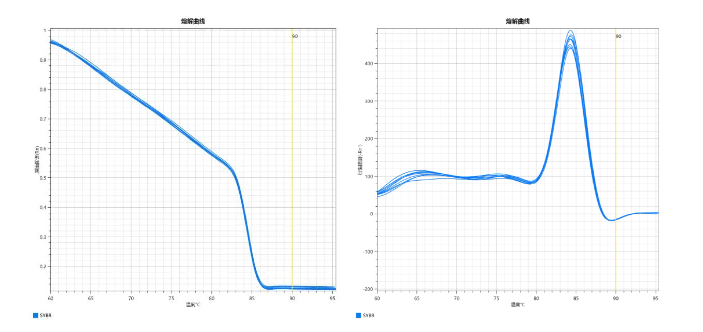
Probe‑Based Melting Curve
Probe‑based melting curve analysis is a technique that uses a specific probe to bind to the target DNA sequence, followed by temperature‑dependent detection of the probe‑target binding status to identify and quantify specific DNA or RNA sequences. The key principle is that when the probe is perfectly matched to the target sequence, its melting temperature (Tm) is higher. The presence of single nucleotide polymorphisms (SNPs) or other variations reduces the Tm. By comparing melting curves of different samples, sequence differences can be accurately identified.
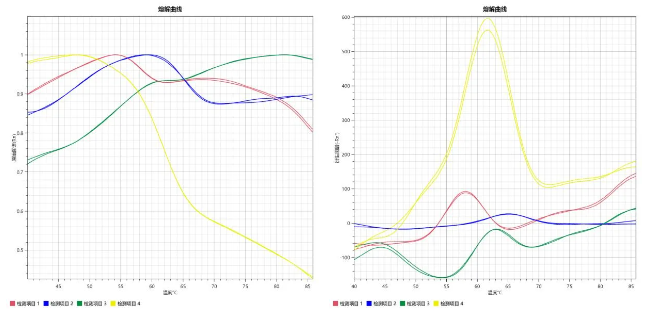
Inverted Melting Curve Peak
Under normal conditions, a melting curve shows a decrease in fluorescence signal as temperature rises. Increasing temperature causes dsDNA denaturation, the fluorescent probe dissociates from the template, and fluorescence decreases, forming a peak from high to low. When the curve shows an inverted peak — where fluorescence increases after a certain temperature — it challenges our understanding. However, this “inverted peak” is not inexplicable. It can be a double‑edged sword: it may indicate non‑specific events, or represent an intentional multiplex detection strategy.
Analysis of Abnormal Inverted Melting Peaks
Probe dissociation kinetics: Probe dissociation from dsDNA can be more complex than expected, especially for certain sequences or conditions. At low annealing temperatures, probes may rebind to alternative sites, causing fluorescence to increase again and forming an inverted peak.
Non-specific products: Additional non‑specific PCR products with different Tm values may cause renewed fluorescence enhancement at higher temperatures.
Primer dimers: Dimers formed between primers may melt at high temperatures, leading to inverted peaks.
Probe design issues: Poorly designed probes may show unstable behavior, such as inappropriate dissociation and rebinding, or secondary structure formation that interferes with template interaction.
Fluorophore properties: Physicochemical changes of some dyes within specific temperature ranges may cause abnormal fluorescence enhancement, such as re‑excitation at high temperatures.
Experimental conditions: Variations in pH, ionic strength, or buffer composition can affect dsDNA stability and probe dissociation, altering melting curve shape.
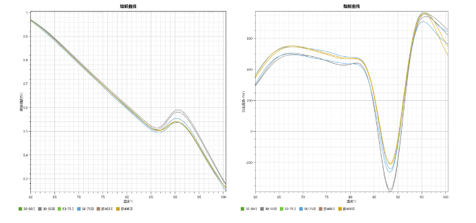
Rational Mechanisms of Inverted Melting Peaks
However, inverted peaks are not always accidental. In advanced experimental designs, carefully engineered probes and conditions allow researchers to use this phenomenon for multiplex target detection. For example, within the same fluorescence channel, one target may show a positive peak while another shows a negative peak. This greatly expands the detection capacity of real‑time PCR, making multiplex assays more efficient and precise.
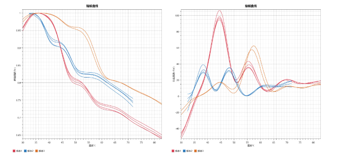
When facing inverted melting curves, researchers should maintain a rigorous scientific attitude, comprehensively reviewing experimental design, PCR conditions, probe and primer characteristics, and instrument calibration. Careful troubleshooting and optimization can correct experimental bias and deepen understanding of the inherent rules of complex biological systems.
Wide Applications, Unlimited Potential
Probe‑based melting curve analysis demonstrates great value across many fields. Using Rocgene’s qPCR analysis software Archimed Analyzer in the Probe Melting Curve module, the following applications can be easily performed:
Disease diagnosis: rapid screening of genetic diseases, infectious pathogens, etc.;
Drug development: evaluating drug effects on specific gene expression;
Food safety: detection of GMO components or pathogenic microorganisms;
Forensic identification: rapid comparison of DNA samples from crime scenes.

The path of scientific research is full of unknowns and challenges. Only with an open mind and rigorous spirit can researchers discover more miracles in the microscopic world. Like the inverted melting curve peak, it is both a puzzle in scientific exploration and a source of inspiration for technological innovation. We must embrace the double‑edged mirror of science: recognize its challenges while appreciating its infinite possibilities. Together, let us explore every corner of science and uncover the mysteries of life sciences.
06
16
08
18


 +86-010-86229225
 info@rocgene.com
 CN
CN